Vertebral Discs Anatomy at Michelle Harris blog Biology Diagrams Intervertebral discs are fibrocartilaginous structures located between the bodies of adjacent vertebrae.They form a fibrocartilaginous joint between the vertebral bodies, linking them together. Collectively, the discs contribute up to one-third of the length of the vertebral column, forming an interpose between adjacent vertebrae from the axis (C2) to the sacrum.
What is an Intervertebral Disc? An intervertebral disc acts as a cushion and spacer between the vertebrae, absorbing shock and allowing flexibility in the spine. Each disc is predominantly composed of two key components: 1. Annulus Fibrosus • The annulus fibrosus is the tough, outer ring of the disc. The intervertebral disc (IVD) is important in the normal functioning of the spine. It is a cushion of fibrocartilage and the principal joint between two vertebrae in the spinal column. There are 23 discs in the human spine: 6 in the cervical region (neck), 12 in the thoracic region (middle back), and 5 in the lumbar region (lower back). Anatomy of the intervertebral disc, the biomechanical properties of the nucleus pulposus and the annulus fibrosus and their responses to a variety of mechanical forces acting on them are discussed. This provides a background for understanding how a healthy intervertebral disc responds to various stresses generated by different movements of the
:max_bytes(150000):strip_icc()/GettyImages-487737771-567da7305f9b586a9eaadecf.jpg)
Intervertebral discs: Anatomy, structure and function Biology Diagrams
Intervertebral discs consist of an outer fibrous ring, the anulus (or annulus) fibrosus disci intervertebralis, which surrounds an inner gel-like center, the nucleus pulposus. [1]The anulus fibrosus consists of several layers (laminae) of fibrocartilage made up of both type I and type II collagen.Type I is concentrated toward the edge of the ring, where it provides greater strength.

Anatomy. An intervertebral disc is one of twenty-four masses of soft tissue found between the vertebral bodies of neighboring vertebrae. Only the space between the atlas (C1 vertebra) and axis (C2 vertebra) lacks an intervertebral disc due to the rotation of the head at the atlantoaxial joint. Each intervertebral disc consists of two main parts

Understanding the Anatomy of an Intervertebral Disc Biology Diagrams
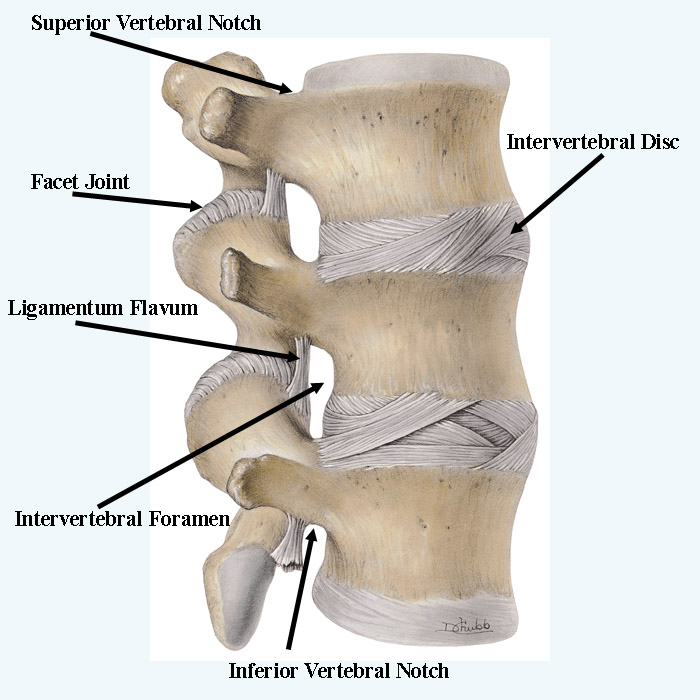
This review article describes anatomy, physiology, pathophysiology and treatment of intervertebral disc. The intervertebral discs lie between the vertebral bodies, linking them together. The components of the disc are nucleus pulposus, annulus fibrosus and cartilagenous end-plates. The blood supply to the disc is only to the cartilagenous end